
Cutting edge publications and research
from the IVF Academy Team.
Embryo Grading: How to Assess Quality and Predict Outcomes
Embryo grading standardizes how labs describe embryo appearance and developmental progress so IVF teams can consistently rank embryos and communicate expectations. It links developmental stage (cleavage vs blastocyst) with morphology to stratify implantation likelihood, which is useful for decision-making, but never a guarantee.
- Most clinics grade at two checkpoints: Day 2/3 cleavage stage (cell number, symmetry, fragmentation, multinucleation) and Day 5/6/7 blastocyst stage (expansion, ICM, TE).
- Cleavage-stage grading is “high signal, high variability,” and works best for ranking embryos within the same cohort.
- Blastocysts are commonly reported as Expansion–ICM–TE (e.g., 4AB): 1–6 for expansion/hatching, then A–C for ICM and TE quality.
- Morphology correlates with implantation/live birth probabilities, but it does not directly measure chromosomal status, endometrial receptivity, or culture conditions, so context and consistency remain essential.
Embryo grading is a structured way to describe embryo appearance and developmental progress. The grading process ensures that lab and clinical teams can consistently rank embryos for transfer or cryopreservation. Likewise, it allows teams to clearly communicate expectations with patients.
Most modern labs follow standardized terminology and scoring conventions to reduce subjectivity and improve reproducibility across observers and clinics. At its core, an embryo grading system links the cleavage stage vs blastocyst and morphology.
With that said, morphology is a proxy. Grading should be used to stratify implantation likelihood and never to “guarantee” outcomes. Still, when applied consistently, IVF embryo grading supports clearer clinical decisions, especially when choosing between multiple embryos.
Continue reading to learn more about how and why IVF practitioners grade embryos.
How Are Embryos Graded in Practice?
The way embryos are graded depends on the day of development and the grading framework your lab uses. International consensus groups (ALPHA/ESHRE) provide definitions for common assessment points and morphologic descriptors, helping labs standardize what they mean by “fragmentation,” “symmetry,” or “blastocyst quality.”
Per the ASRM, most programs assess embryos at two major checkpoints:
- Cleavage stage (Day 2/3): focuses on cell number and early morphologic markers.
- Blastocyst stage (Days 5/6/7): focuses on expansion, inner cell mass, and trophectoderm features, often using the widely adopted Gardner-style approach.
These checkpoints help teams communicate embryo grades in a standardized way while acknowledging that embryos can change meaningfully over time.
Cleavage-stage Embryo Grades: What Matters and Why
Cleavage-stage assessment is often described as “high signal, high variability.” It can be informative within a cohort, but it’s also more vulnerable to timing differences and observer interpretation.
From a practical standpoint, cleavage-stage embryo grades generally incorporate:
- Cell number relative to expected timing (developmental pace)
- Blastomere symmetry (more even cleavage can be favorable)
- Fragmentation percentage (higher fragmentation is typically less favorable)
- Multinucleation (often associated with poorer prognosis)
Rather than over-weighting any single feature, many labs use cleavage-stage grading to rank embryos within the same cycle. From there, they can refine selection if embryos progress to blastocyst. That’s because blastocyst development provides additional structure-based markers and an added developmental “filter.”
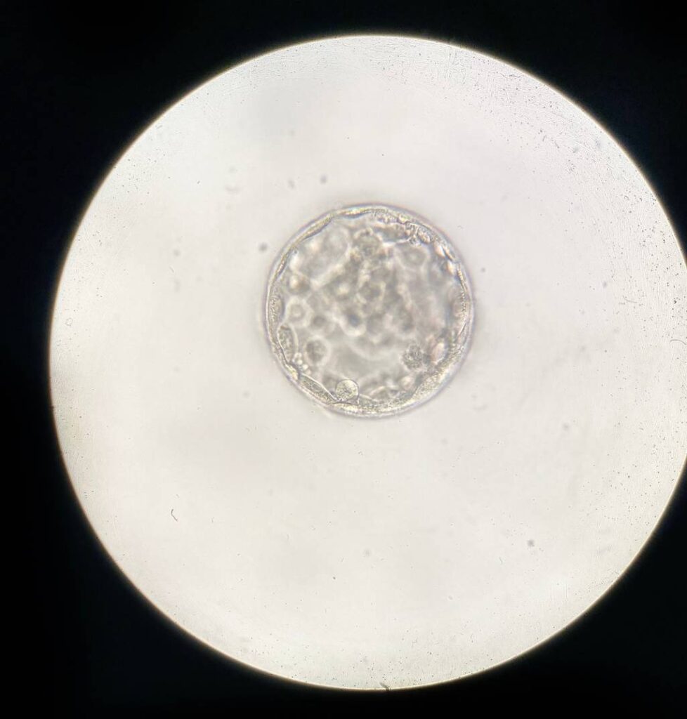
Blastocyst Grading: Expansion, ICM, and TE
Blastocyst grading tends to be more clinically intuitive because it aligns with key embryo structures and their future roles.
A common framework describes the blastocyst using:
- Expansion stage (how expanded/hatching the blastocyst is)
- Inner Cell Mass (ICM) quality (cells that form the fetus)
- Trophectoderm (TE) quality (cells that contribute to placental tissues)
Blastocysts are typically reported as Expansion–ICM–TE. For example, “4AB.”
The number (1–6) describes the degree of blastocoel expansion and hatching status (with higher numbers indicating a more expanded or hatching/hatched blastocyst). The first letter grades the inner cell mass (ICM). The second letter grades the trophectoderm (TE), commonly A (many, cohesive cells), B (moderate/less cohesive), or C (few/irregular cells).
What Morphology is Really Telling You About Embryo Quality
When clinicians or embryologists talk about embryo quality, they’re often referring to the embryo’s apparent developmental competence based on morphology and timing. Evidence across studies suggests that blastocyst morphologic parameters, especially TE and ICM characteristics, are associated with implantation and live birth probabilities. This is despite the fact that the relative weight of each component can vary by population and study design.
A useful way to frame this clinically is to say that blastocyst grading is probabilistic triage. It helps decide which embryo to transfer first and which to prioritize for cryopreservation. However, it does not replace genetic information or patient-specific factors.
There are many things that embryo grade can’t predict. Even strong-looking embryos can be aneuploid, and lower-graded embryos can result in healthy live births. That’s why consensus guidance stresses consistency and context, not over-interpretation.
Embryo morphology does not directly measure:
- chromosomal status
- endometrial receptivity and synchrony
- culture conditions across incubators/media
- underlying oocyte/sperm contributions beyond what’s visible
While embryo grading supports decision-making, the best clinical use is as part of an integrated selection strategy.
Optimizing the IVF Outcomes Equation
IVF embryo grading helps teams communicate clearly and make defensible choices. Used well, it connects visible morphology and developmental progression to the likelihoods of implantation and live birth while staying grounded in the reality that embryo grades are only one part of a much bigger equation.
To strengthen consistency in embryo assessment and lab-to-clinic communication, ongoing training matters. Learn more about laboratory courses at IVF Academy.